
Andy Chervenak, Graduate Student, Department of Cell and Developmental Biology, University of Michigan Medical School
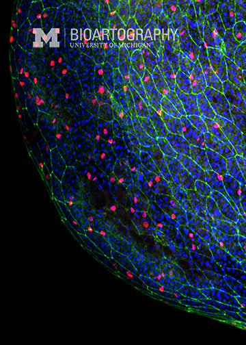
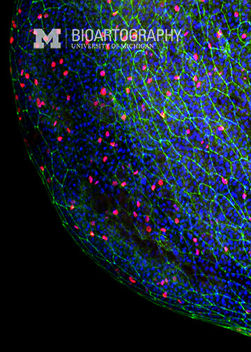
This is an embryo of a Zebrafish (Danio rerio), a tropical freshwater fish related to the minnow family. Individual cells on the surface of the embryo are marked by staining an adhesion protein (E-cadherin, green) that glues the cells together. The red dots are nuclei that have just finished synthesizing DNA in preparation for mitosis. The Zebrafish embryo is a widely used model organism for developmental biology studies, since the embryos are transparent and develop outside of the body. Study of the development of the zebrafish embryo has provided important insight into the development of many human organs, including the heart, muscles, eye, gut and brain.
