
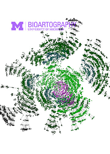
In developing vertebrates, discrete round groups of cells called somites form on both sides of the central neural tube on the back of the embryo. The somites are transient structures whose cells migrate great distances to give rise to the vertebrae and ribs, the muscles of the back and limbs and the skin of the back. Interestingly, somites form according to an internal clock; in the chick, one somite is formed every 90 minutes. This network illustrates a time-course (over 90 minutes) of interactions among 13 segmentation clock genes (and proteins) responsible for the formation of the precursors of vertebrae in the chick. Each dot represents the expression of a gene (or protein), and the gray lines represent interactions among the genes/proteins. Initial states are on the periphery and the stable and long-term state of interactions among these genes is illustrated at the center by the heavier black line. The change in the colors of the dots indicates the evolution of gene/protein expression towards the stable pattern that leads to the periodic formation of vertebrae precursors during embryo development.