
Benjamin Carlson, Graduate Student, Cell Biology, Duke University
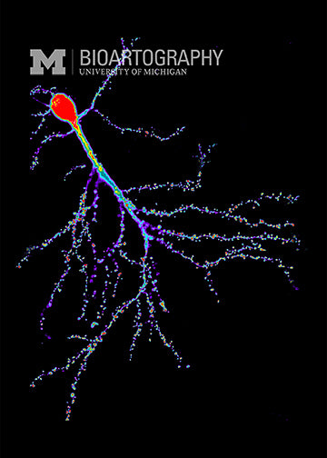
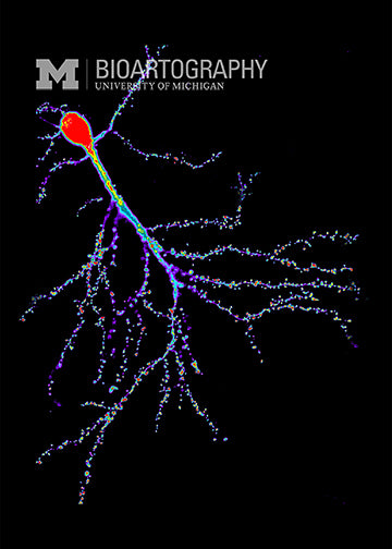
This image is a mouse neuron from the hippocampus, the brain area that is critically involved in memory. The large branching projections contain spiky spines at their tips that receive signals from other neurons. These spiny structures are thought to be involved in the processing of information during learning and memory function. In this image, the neuron is stained to identify a protein that forms the internal skeleton of the cell. This allows us to study the structure of these spines during memory formation and learning.
