

Ann Grosse, Graduate Student, Department of Cellular and Molecular Biology, University of Michigan Medical School
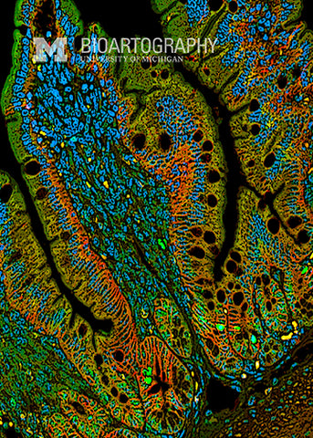
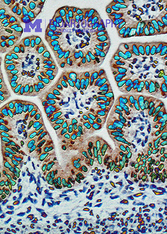
The luminal surface of the intestine is highly convoluted to increase its surface area for absorption. This convolution is provided by billions of finger-like structures called villi (singular, villus). Villus development occurs during embryonic life. This is an image of the fetal mouse intestine during the time that villi are just starting to be organized. The membranes of the cells are stained red and yellow; the dense packing of the developing villi is obvious. Little is understood at the molecular level about how the villi are formed and maintained. This research will be important to find better treatments for adults with the reduced intestinal surface area after bowel resection or newborn infants with short bowel syndrome.
