

Åsa Kolterud, Ph.D., Postdoctoral Fellow, Department of Cell and Developmental Biology, University of Michigan Medical School
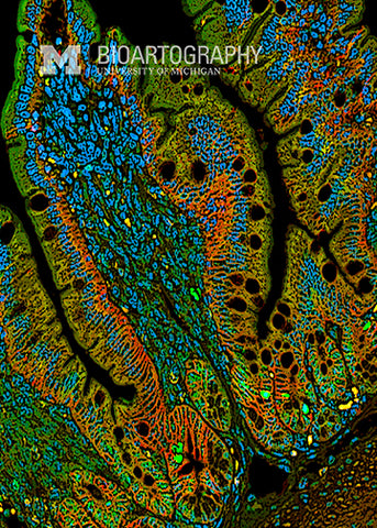
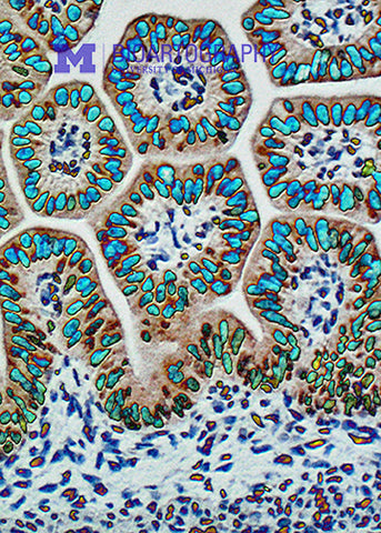
During embryogenesis, the developing intestine undergoes a remarkable remodeling process in which the surface of the gut tube is folded into finger-like projections called villi. These villi, which extend into the lumen of the gut tube, drastically increase the absorptive surface of the intestine and are important for efficient nutrient uptake. This photograph shows the initial buckling of the intestinal epithelium (stained red) into nascent villi. The nuclei of the cells are stained blue. Note the flower-like nuclei within the epithelium – these are dividing cells lining up their chromosomes.
