

Ann Grosse, Graduate Student, Department of Cellular and Molecular Biology, University of Michigan Medical School
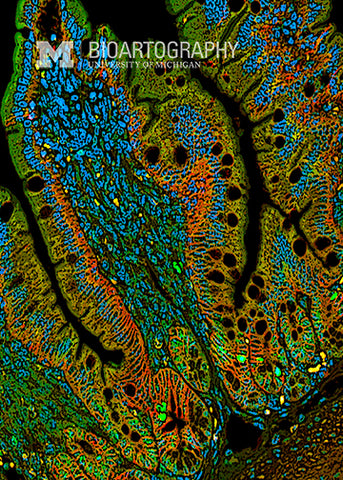
The inside of the gut is lined with millions of finger-like projections called villi (singular, villus) that increase the surface area and maximize the absorptive capacity of the intestine. This image is a cross-section of the fetal mouse intestine highlighting the structure, pattern, and organization of the villi. Each villus is outlined in red by a marker that recognizes the luminal surface of the epithelial cells. Nuclei are stained blue with a DNA marker, and blood cells are yellow. Nutrients are absorbed in the apical surface, travel through the epithelium, enter the bloodstream, and are delivered throughout the body. Disruption of the intestinal lining from disorders such as celiac disease or inflammatory bowel disease can result in distortion of the villi and contribute to the development of malabsorption.
