
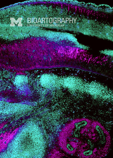
Ilea Swinehart, Graduate Student, Cellular & Molecular Biology Program, University of Michigan Medical School
This image is a longitudinal section of a mouse embryo. At the top, is the neural tube, or the developing spinal cord. The segmented balls of tissue underneath of are the somites, the tissue that gives rise to the vertebrae, the muscles and the dermal layer of the skin. The early developing kidney can be seen in the bottom right of the photograph. By mutating mouse DNA, we can introduce changes into genes that we believe might control the development of these organs. Then, sections such as these are used to determine which organs are affected and how. The changes we see in the mouse very often mirror similar developmental birth defects seen in humans.