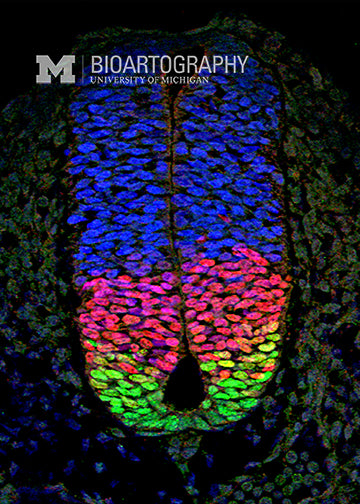
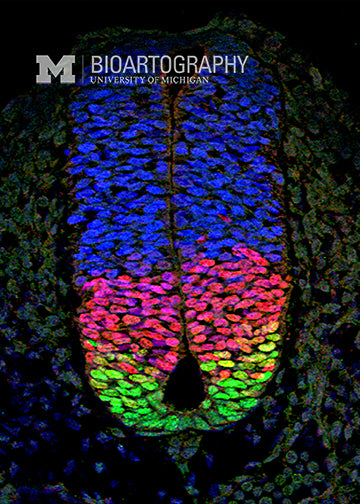
This image depicts a transverse section through the spinal cord of a developing mouse embryo (embryonic day 9.5 of development; the normal gestation period for mice is 21 days). The dorsal (back) side of the embryo is at the top; the ventral (belly) side is at the bottom. The different colors represent distinct groups of nuclei that comprise different neural progenitor populations. These patterns are defined by precisely controlled cell fate decisions during embryogenesis. The green cells denote interneurons that connect different populations of neurons that regulate locomotion. The red cells mark motor neurons that control voluntary and involuntary movements. Finally, the blue nuclei represent more dorsal neurons that process sensory information. The cell fate decisions that are depicted in this snapshot of a relatively early stage of spinal cord patterning are essential for proper brain and neuronal development.

