
Kate Walton, Ph.D., Research Assistant Professor (Gumucio Laboratory), Department of Cell and Developmental Biology, University of Michigan Medical School
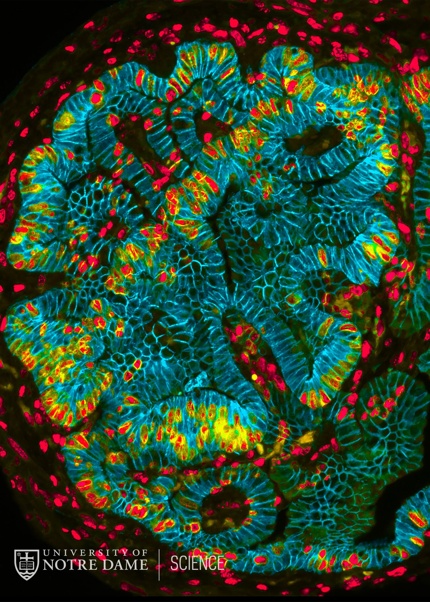
The lumen of the small intestine is studded with finger-like projections called villi that serve to increase the surface area available for absorption of nutrients. This image shows the rapidly growing absorptive surface of a fetal mouse. The red nuclei are stained for a marker of dividing cells so you can see how actively the tissue is growing! The cells outlined in blue will be in contact with the food supply and gut microbes; they function to absorb nutrients but also act as a barrier to keep the bacteria inside the gut lumen from entering the body.
18-034