Sha Wang, Ph.D., Postdoctoral Fellow (Gumucio Laboratory), Department of Cell and Developmental Biology, University of Michigan Medical School
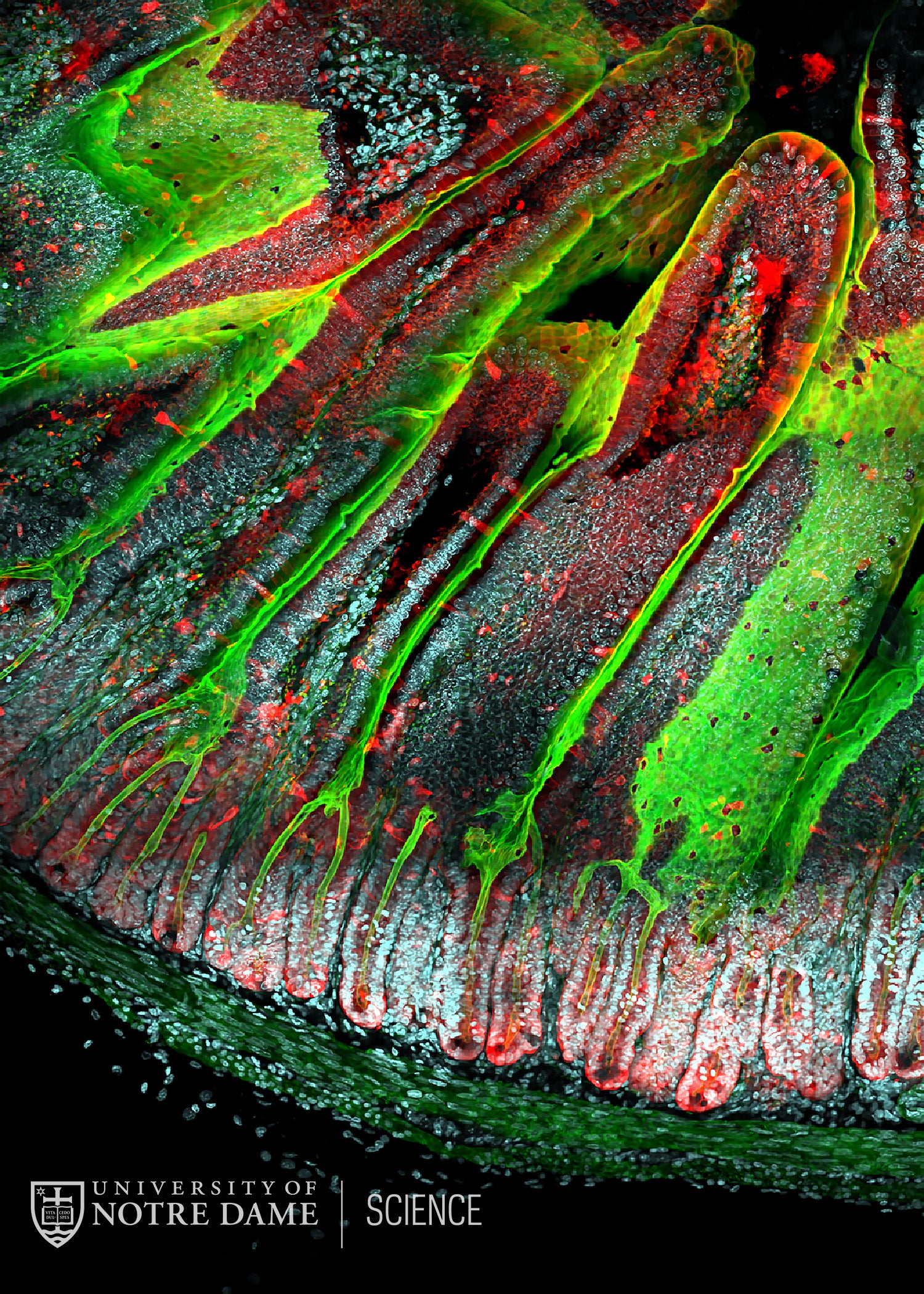
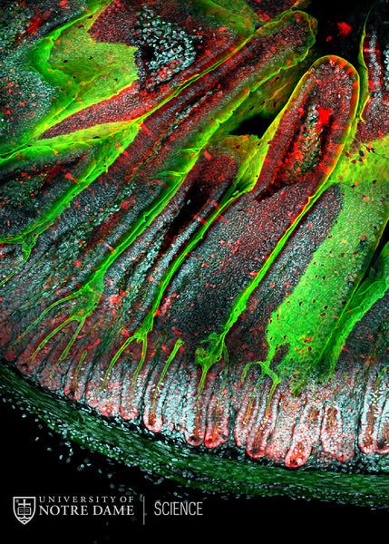
The lining of the small intestine is specialized to have an enormous surface area that allows it to efficiently absorb nutrients from ingested food. In this image from a 3-week old mouse, all of the epithelial cells (which form the intestinal lining) are labeled in red. The surface of the epithelial cells is marked in green, highlighting the fact that the epithelial surface is thrown into finger-like projections called villi, which serve to greatly amplify the available surface area. The epithelium itself is made of a very dynamic population of epithelial cells. The cells on the tops of the villi are regularly shed and lost into the intestinal lumen; meanwhile, a group of stem cells resides deep in the bottom of the crypts (tube-like structures at the bottom of the image). The stem cells divide rapidly to offset the loss of cells at the villus tips, renewing the entire epithelial cell population every 4-5 days.
22-083