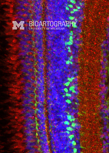

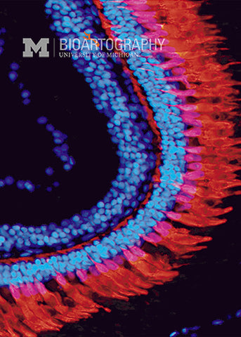
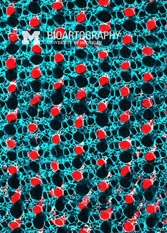
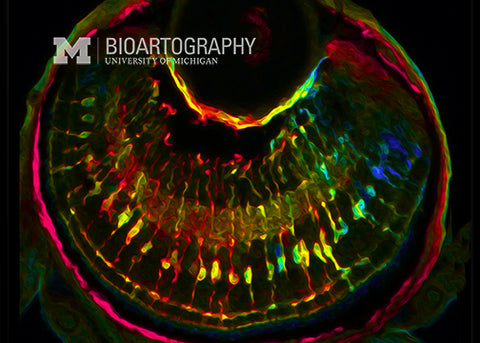
Jenny Lenkowski, Ph.D., Postdoctoral Fellow, Department of Molecular, Cellular and Developmental Biology, University of Michigan
This image is a cross-section of an adult zebrafish retina. The blue dots in the image mark cell nuclei, the red stain detects a protein called TGIF, which is an important regulator of retinal cell regeneration. The green cells are Müller glia. Glial cells were originally thought to serve only support functions, but recent data indicate that they can give rise to multipotent progenitor cells. Our laboratory is exploring how Müller glia know how to make cone photoreceptors to regenerate a damaged retina, as well as the role that the TGIF protein may have in this regeneration process.