
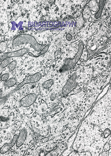
This picture was taken on the electron microscope. Using electrons, scientists are able to image the inside of cells in great detail. This micrograph shows the region of the cell near the nucleus. The grey elongated structures in the center are mitochondria, the energy factories of the cell. The nucleus, which holds the DNA, is at the bottom right. In this micrograph, there are several abnormal blebs in the membrane surrounding the nucleus. This is due to a genetic mutation in a gene called TorsinA that causes the childhood disease DYT1 dystonia, a debilitating movement disorder in which children exhibit involuntary twisting motions. The amazing feature of this disease is that while TorsinA is expressed by many cells of the body, only neurons exhibit abnormal nuclear membranes.
