

Nikolas Kazmers, Undergraduate Student, Department of Engineering, University of Michigan
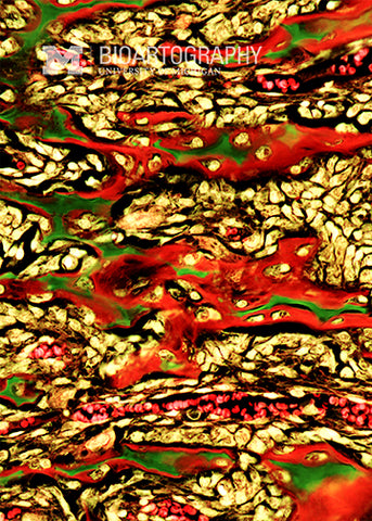
Mouse bone cells wander along the glass surface of a tissue culture dish. The cells are stained for structural proteins called vinculin (green) and actin (red). Vinculin is a protein present in specialized regions of the cell that allow it to attach to a surface. These transient suction cups (green patches) allow a cell to generate the force needed to move. Inside the cell, these focal adhesions are connected to a protein called actin (red), which forms long cables that act like supporting guy wires to give the cell its shape and provide structural integrity.
