
Esther Miranda, Research Analyst, Cell Biology, Duke University
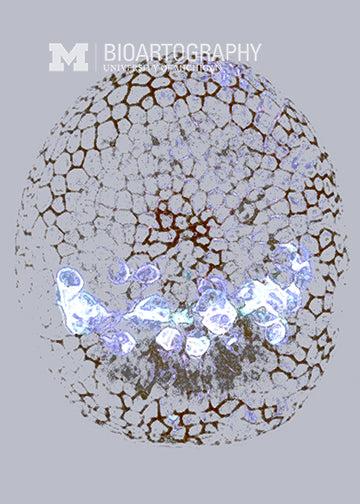
This sea urchin embryo was stained to identify the primary mesenchyme cells (white) that will later generate the skeleton of the sea urchin. These cells arise from the outer layer of cells (stained black) and migrate in a characteristic way to the inside of the embryo. These primary mesenchyme cells are used by researchers as a model to study cell migration and its involvement in embryonic development and cancer metastasis.